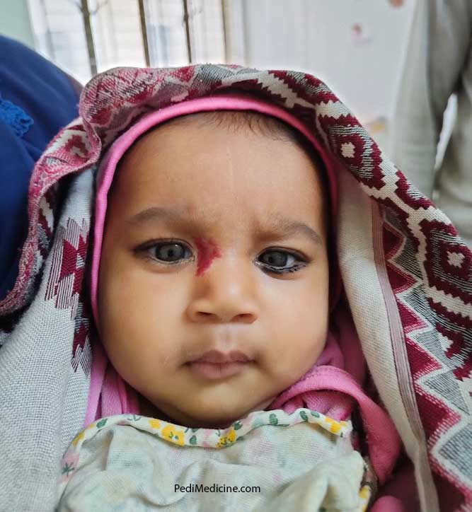
1. What is Congenital Hemangioma?
Hemangioma is a benign tumour. Benign means it’s not that dangerous. So there is not much to worry about. It is a benign tumour of blood vessels. A hemangioma that presents since birth is called a congenital hemangioma. It is fully formed at birth. Blood vessels are formed by the cells known as endothelial cells. When they grow more than they should, they are called a hemangioma. The extra tissue that is formed is now attached to the normal vessels. Congenital hemangioma comes in many shapes. It is usually round or oval or may be irregular. They may be reddish to pinkish or may be bluish in colour. They are fully grown when the baby is born and do not grow after birth. they usually shrink away. Congenital hemangioma usually does not need treatment. If it does not shrink away or present in a vital area of the body your doctor will suggest the necessary treatment. If it is causing any problem usually surgery is done. Laser treatment is also available.
2. What is Infantile Hemangioma?
Infantile hemangioma usually appears at birth or later in the first few weeks of life. They grow rapidly for the first few months of life. Mostly they stop growing at a certain age and later involute. Infantile hemangioma is the most common tumour that affects babies. It usually disappears at 4-5 years of life and leaves behind scars or extra blood vessels on the skin. The skin may be slightly discoloured or raised after the hemangioma goes away. Infantile hemangiomas are much more common than congenital hemangiomas.
List of Common Childhood Cancers and Tumors
3. Why does Hemangioma occur?
The exact cause is unknown. Infantile hemangiomas are more common to occur in girls than boys and are more common in Caucasian children. Babies who are born early or who have low birth weight are more likely to have an infantile hemangioma.

4. Types of infantile hemangioma
In most cases, the infantile hemangioma is superficial and bright red in colour. Sometimes they are called strawberry birthmarks. They are known as superficial hemangioma.
If they are deep under the skin and look either bluish or skin-coloured; these are called deep infantile hemangiomas.
When both are present simultaneously they are called a mixed infantile hemangioma.
5. How to diagnose infantile hemangiomas
Infantile hemangioma is diagnosed by just looking at it. No special test is necessary. But if it is very large and involves vital organs or areas an Ultrasonogram or MRI can be done. MRI is done to see it involves other organ systems. Sometimes a hemangioma is a part of PHACE syndrome. PHACE stands for
- P= Posterior fossa malformation.
- H= Hemangioma
- A= Abnormal arteries in the brain or near the heart
- C= Coarctation of the aorta
- E= Eye involvement
6. How to treat infantile hemangioma
Hemangiomas are usually treated by a paediatrician, a dermatologist and sometimes a haematologist or a cosmetic surgeon if it needs surgery. Mostly it does not need any treatment. Observation is necessary to see if it’s increasing in size rapidly. Treatment depends upon the fact that how large it is, or does it involve a major organ system or cause any problem. Sometimes the cosmetic cause is the reason to so surgery.
A Beta-blocker, propranolol is approved for the treatment of infantile hemangioma by FDA. [Source]
Timolol can also be used for treating hemangioma. Timolol drop is available. It can be applied directly to the hemangioma surface of the skin. It is used to treat smaller, thin, infantile hemangiomas.
Surgery is less common nowadays. Most surgeries for hemangiomas can be done as outpatient procedures. This means that children can go home the same day they have surgery. When surgery is needed, it is usually done before school age to repair damage or scars caused by infantile hemangioma.
if your baby has rashes on the skin see here “Baby Rashes Treatment and Prevention”
7. Where does it commonly occur?
Around the eye, face, Lips, the upper part of the chest, mouth, genital area, scalp, back etc. Hemangiomas also may develop in organs inside the body, such as the kidneys, lungs, liver, or brain, where they can’t be seen.
Summary review by PubMed Infantile Hemangioma: An Updated Review
The majority of infantile hemangiomas are not present at birth. They often appear in the first few weeks of life as areas of pallor, followed by telangiectatic or faint red patches. Then, they grow rapidly in the first 3 to 6 months of life. Superficial lesions are bright red, protuberant, bosselated, or with a smooth surface, and sharply demarcated. Deep lesions are bluish and dome-shaped. Infantile hemangiomas continue to grow until 9 to 12 months of age, at which time the growth rate slows down to parallel the growth of the child. Involution typically begins by the time the child is a year old. Approximately 50% of infantile hemangiomas will show complete involution by the time a child reaches age 5; 70% will have disappeared by age 7; and 95% will have regressed by 10 to 12 years of age. The majority of infantile hemangiomas require no treatment. Treatment options include oral propranolol, topical timolol, and oral corticosteroids. Indications for active intervention include haemorrhage unresponsive to treatment, impending ulceration in areas where serious complications might ensue, interference with vital structures, life- or function-threatening complications, and significant disfigurement.
Further Reading:
- Mayoclinic Hemangioma
- www.hemangiomaeducation.org
- Wikipedia Hemangioma
- Infantile haemangioma: Definition and pathogenesis Dermnetnz.org
ICD Code for Infantile Hemagioma: D18. 01 – Hemangioma of skin and subcutaneous tissue.


